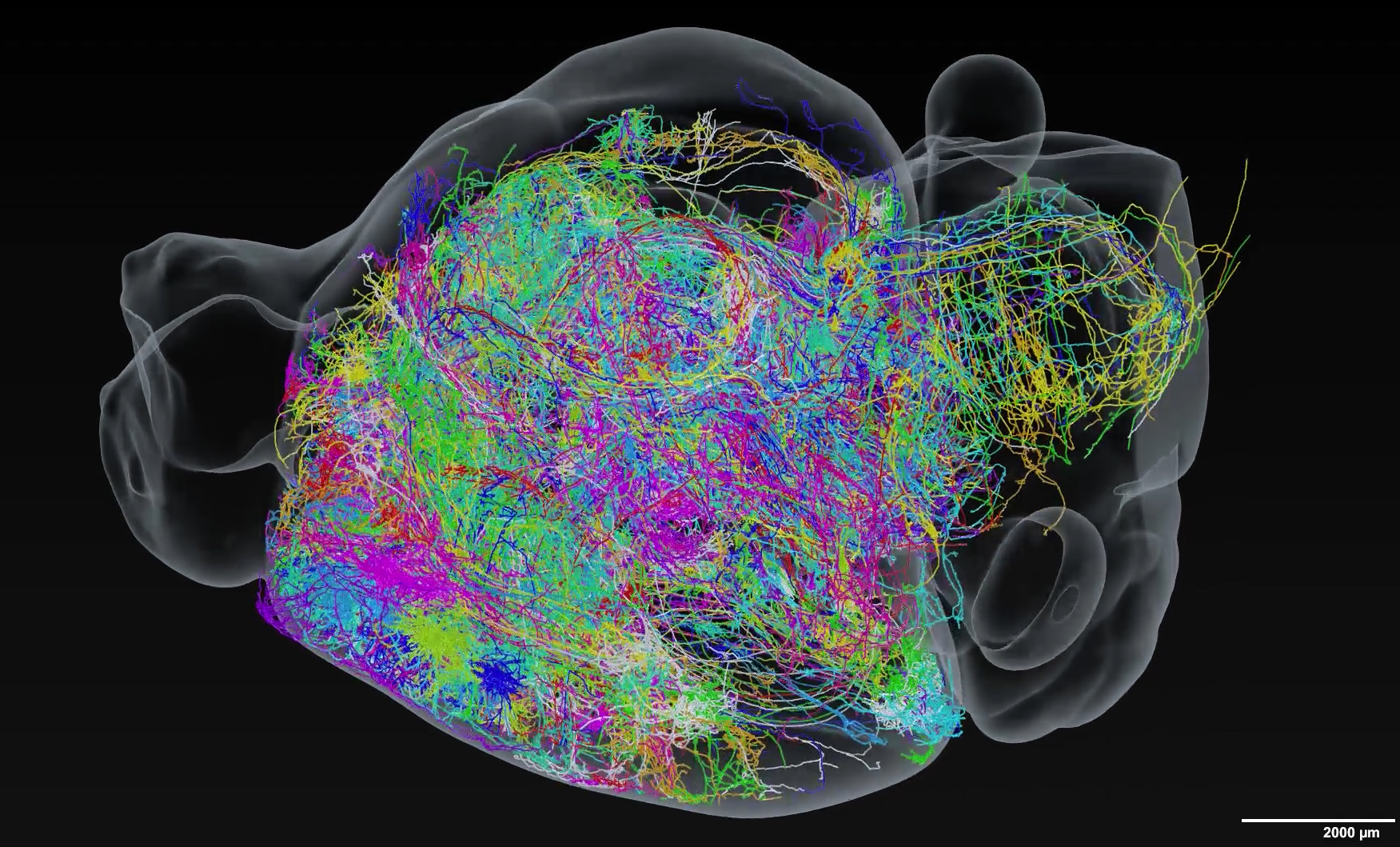
You can now visualise individual neurons firing and zigzagging across the brain in this incredible series of cerebral maps.
Created by researchers from the Janelia Research Campus at the Howard Hughes Medical Institute, the 3D maps represent the most extensive network of brain wiring ever made.
Beyond teaching us more about the structure of the brain, these maps could finally help unlock some of the secrets of memory, taste, touch and more in mice, and in humans.
Scroll down to see neurons firing in 3D or visit the MouseLight NeuronBrowser to explore specific brain regions
Previous attempts to trace the paths of individual neurons have been limited – mapping around a dozen neurons at a time – because the process involves analysing millions of images for each neuron.
Specifically, scientists have to light up neurons inside the brain, slice it into thin sheets, image the sheets with a microscope, and then highlight single neurons among the millions of images collected. By fine-tuning each step of this process, the researchers from Janelia Research Campus were able to reconstruct the entire shape and position of more than 300 of the roughly 70 million neurons in the mouse brain.
This involved injecting mouse brains with a virus that highlights a few dozen neurons. The team then “cleared” the brain, to help light penetrate the tissue, before a high-tech light microscope hit the brain with pulses of light. These pulses highlighted neurons and were used to shave 200-micrometer brain slices with a vibrating razor blade. The whole process was then repeated until the entire brain was imaged.
Each brain produced around 20 terabytes of data – about the storage space of 4,000 DVDs – before algorithms were used to stitch the images together. A team of seven trained individuals then scoured the resulting dataset to digitally untangle individual neurons.
“These people are full-time neuron tracers; their efforts and the software they use are critical for high-quality mapmaking”, said neuroscientist Nelson Spruston, the senior director of scientific programs at Janelia.
“Two years ago, each person would have needed a week or two to track a neuron’s path through the brain. Today, by using neuron-tracing algorithms and software developed by MouseLight and Janelia’s Scientific Computing group, each team member can track roughly one neuron per day.”
The work is significant because we know very little, relatively, about how neurons transmit messages acorss the brain. Typically, neurons are depicted as “fried eggs with tails” and as they send messages through these tails, known as axons, neighbouring neurons pick up the messages via branching tendrils called dendrites. However, this view is largely oversimplified and more data is needed to know exactly how these messages are sent, received and what can cause these networks to degrade.
From their analysis of these 300 neurons the researchers have already discovered neurons are often vastly more far-reaching and connected than once thought. For example, the axons of individual neurons in the thalamus often branch into unexpected combinations of cortical areas, such as those involved in taste, touch, and movement.
Similarly, in the subiculum, a region linked to learning and memory, neurons almost always reach out to a few different places. One neuron the researchers traced ran scattershot across the cerebral cortex, sending long, branching axons arcing across both hemispheres like a “fireworks explosion”. By understanding how these neurons are connected, the researchers are not only unlocking secrets about how memories form, as an example, they could also reveal why some people taste food differently to others, and how diseases such as Alzheimer’s can impact brain function.
“Three hundred neurons is just the start,” said neuroscientist Jayaram Chandrashekar, who leads the Janelia MouseLight team. He and colleagues expect to trace hundreds more neurons in the coming months.
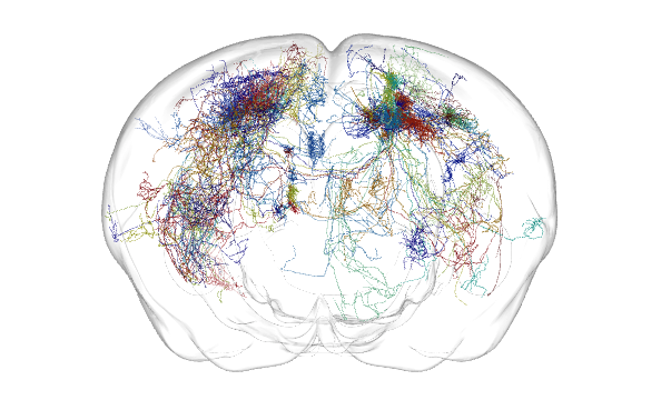
Neuroscientist Eve Marder of Brandeis University and Janelia member added: “We knew that different regions of the brain talked to each other, but seeing it in 3-D is different. The results are so stunning because they give you a really clear view of how the whole brain is connected.”
The team reported the release of their current dataset and an analysis tool, called the MouseLight NeuronBrowser, on November 13, 2017 at the annual Society for Neuroscience meeting in Washington, D.C.
Images/Video: Janelia Research Campus, MouseLight project team
Disclaimer: Some pages on this site may include an affiliate link. This does not effect our editorial in any way.